Oct 30, 2023A tissue is a group of cells, in close proximity, organized to perform one or more specific functions. There are four basic tissue types defined by their morphology and function: epithelial tissue, connective tissue, muscle tissue, and nervous tissue. Epithelial tissue creates protective boundaries and is involved in the diffusion of ions and
Fascia, Bones, and Muscles – BeingHuman
Nov 3, 2023It matures into other types of connective tissues, muscles, vessels, mesothelium and the urogenital system. Its mesenchymal cells are dispersed within ECM filled mainly with reticular fibers. Mucoid connective tissue is found in the umbilical cord. Its mesenchymal cells are loosely distributed within a collagen rich ECM called Wharton’s jelly.
Source Image: chegg.com
Download Image
Unlike cardiac and smooth muscle, the only way to functionally contract a skeletal muscle is through signaling from the nervous system. Figure 2.7.1 2.7. 1 : The Three Connective Tissue Layers Bundles of muscle fibers, called fascicles, are covered by the perimysium. Muscle fibers are covered by the endomysium.

Source Image: m.facebook.com
Download Image
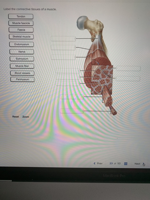
Microscope World Blog: Tendons under Microscope The activity linked below is a drag and drop activity for students to practice labeling the muscles, there are 6 slides showing images of muscles and fibers and the connective tissue surrounding the fibers (endomysium, perimysium, epimysium). Drag and drop activity for remote learners to practice labeling muscles, focusing on the cells and

Source Image: histologydrawings.blogspot.com
Download Image
Label The Connective Tissues Of A Muscle.
The activity linked below is a drag and drop activity for students to practice labeling the muscles, there are 6 slides showing images of muscles and fibers and the connective tissue surrounding the fibers (endomysium, perimysium, epimysium). Drag and drop activity for remote learners to practice labeling muscles, focusing on the cells and The general structure of intramuscular connective tissue. (A) Schematic diagram showing the general arrangement of the epimysium, perimysium, and endomysium within muscle.(B) Schematic diagram depicting the sparse junction zones between the thick perimysium and the endomysium of muscle fibers in the surface layer of the fascicle.(C) Schematic diagram showing myofibrils of an individual muscle
Muscle Tissue
These and other connective tissues associated with muscles follow: The endomysium is the connective tissue that surrounds each muscle fiber (cell). The perimysium encircles a group of muscle fibers, forming a fascicle. The epimysium encircles all the fascicles to form a complete muscle. A tendon is a cordlike extension of the preceding three Tissue – Definition, Meaning, Types of Tissue

Source Image: medilogbiohealth.com
Download Image
Fauces – pediagenosis These and other connective tissues associated with muscles follow: The endomysium is the connective tissue that surrounds each muscle fiber (cell). The perimysium encircles a group of muscle fibers, forming a fascicle. The epimysium encircles all the fascicles to form a complete muscle. A tendon is a cordlike extension of the preceding three

Source Image: pediagenosis.com
Download Image
Fascia, Bones, and Muscles – BeingHuman Oct 30, 2023A tissue is a group of cells, in close proximity, organized to perform one or more specific functions. There are four basic tissue types defined by their morphology and function: epithelial tissue, connective tissue, muscle tissue, and nervous tissue. Epithelial tissue creates protective boundaries and is involved in the diffusion of ions and
Source Image: magicalrobot.org
Download Image
Microscope World Blog: Tendons under Microscope Unlike cardiac and smooth muscle, the only way to functionally contract a skeletal muscle is through signaling from the nervous system. Figure 2.7.1 2.7. 1 : The Three Connective Tissue Layers Bundles of muscle fibers, called fascicles, are covered by the perimysium. Muscle fibers are covered by the endomysium.
Source Image: blog.microscopeworld.com
Download Image
Multitier mechanics control stromal adaptations in the swelling lymph node | Nature Immunology Oct 30, 2023Regardless of its morphology or type, muscle tissue is composed of specialized cells known as muscle cells or myocytes (myo- [muscle, Greek = mys]), commonly referred to as muscle fibers (all of these terms are interchangeable); this is due to their extensive length and appearance. Myocytes are characterized by protein filaments known as actin and myosin that slide past one another, producing

Source Image: nature.com
Download Image
Importance Of Foot And Ankle Strengthening For Dancers The activity linked below is a drag and drop activity for students to practice labeling the muscles, there are 6 slides showing images of muscles and fibers and the connective tissue surrounding the fibers (endomysium, perimysium, epimysium). Drag and drop activity for remote learners to practice labeling muscles, focusing on the cells and

Source Image: yumpu.com
Download Image
This blogger has esophageal cancer – HoCoMDcc The general structure of intramuscular connective tissue. (A) Schematic diagram showing the general arrangement of the epimysium, perimysium, and endomysium within muscle.(B) Schematic diagram depicting the sparse junction zones between the thick perimysium and the endomysium of muscle fibers in the surface layer of the fascicle.(C) Schematic diagram showing myofibrils of an individual muscle

Source Image: hocomd.cc
Download Image
Fauces – pediagenosis
This blogger has esophageal cancer – HoCoMDcc Nov 3, 2023It matures into other types of connective tissues, muscles, vessels, mesothelium and the urogenital system. Its mesenchymal cells are dispersed within ECM filled mainly with reticular fibers. Mucoid connective tissue is found in the umbilical cord. Its mesenchymal cells are loosely distributed within a collagen rich ECM called Wharton’s jelly.
Microscope World Blog: Tendons under Microscope Importance Of Foot And Ankle Strengthening For Dancers Oct 30, 2023Regardless of its morphology or type, muscle tissue is composed of specialized cells known as muscle cells or myocytes (myo- [muscle, Greek = mys]), commonly referred to as muscle fibers (all of these terms are interchangeable); this is due to their extensive length and appearance. Myocytes are characterized by protein filaments known as actin and myosin that slide past one another, producing